
Diagnostic imaging
MRI, CT, Nuclear scintigraphy, radiography, ultrasound and standing CT.
Magnetic Resonance Imaging (MRI)
MRI is an excellent tool for evaluating the structures within the foot, and we also have experience of using it for image structures up to and including the carpus and tarsus.
MRI scans are performed by our qualified equine veterinary nurses, who also have training in equine diagnostic imaging and can obtain the best quality images possible.
The use of MRI has revolutionised our approach to foot lameness and penetrating injuries of the foot with a definitive diagnosis being achieved in 90% of cases. The use of MRI can save huge amounts of time and money that can be wasted in the absence of a definitive diagnosis, and then allows for specific treatments to be undertaken. MRI should be considered early in cases of foot lameness that cannot be diagnosed by traditional methods.
MRI scans are performed standing under sedation and do not require a general anaesthetic. Further information on MRI and its uses in equine medicine is available at www.hallmarq.net. Our referral team, led by Jonathon Dixon BVetMed MVetMed DipECVDI MRCVS are always happy to discuss cases that you think may benefit from an MRI scan and can be contacted on imaging@rainbowequinehospital.co.uk for further advice.
For more information on what an MRI scan entails see admission for MRI.
Computed tomography (CT)
Our CT scanner has a wide bore, allowing us to fit larger regions of our patients in the scanner, improve image quality and enable scans to be completed in only 30 seconds.
CT is the acquisition of a series of x-ray images at high speed circumferentially around the area of interest so giving a 3-dimensional x-ray image. It has significant advantages over 2-dimensional x-ray particularly in the assessment of dental, sinus and other head diseases and in the assessment of fractures.
In adult horses we are able to image the head and proximal neck with the horse standing up and sedated, allowing us to accurately assess the teeth and paranasal sinuses, and the skull bones. These scans are done with the horse floating on a stable bed of air, and are both rapid and completely non-invasive for your horse. This is the gold standard imaging test for the diagnosis of dental disease and we would strongly recommend its use in cases where oral examination and oroscopy has been unable to determine a definitive diagnosis.
In horses under general anaesthesia (supervised by Kate Loomes BVSc CertAVP(EP) CertAVP (VA) DipECVAA MRCVS, Specialist in Veterinary Anaesthesia), we can image the entire neck (cervical spine) to the cranial thoracic region, including performing myelograms. For lameness and limb issues, we can scan from the foot to the elbow in the forelimb and the foot to stifle in the hindlimb, with pelvic CT possible in some patients too; this has completely revolutionised equine imaging and we are proud to be able to offer this. We frequently complement limb imaging with use of an x-ray contrast media with this either injected into a joint to delineate the articular cartilage, or using ultrasound to inject this into an artery to highlight soft tissue injuries.
Recently the scanner capabilities have allowed us to begin to perform thoracic and abdominal CT examinations in foals and smaller horses and ponies, pushing back the boundaries of technology to achieve the best for our patients, this can be used to evaluate the blood vessels (angiography), kidneys and intestinal tract amongst other things, though in some of our largest horses the size may still limit what can be included.
Our referral team are always happy to discuss cases that you think may benefit from a CT scan and for more information on what a CT scan entails see admission for CT.
Nuclear scintigraphy (bone scan)
We have the latest scintigraphy equipment to provide the best possible images.
Following the horse being exercised carefully and the subsequent injection of a radiopharmaceutical medication (99mTc-HDP), a gamma camera is used to detect areas of increased bone activity or “hot spots”.
Horses undergoing a bone scan must remain in the hospital for 48-hours after the injection, as they will still be radioactive.
Increased bone activity is expected where there has been injury, damage or joint disease and the bone is trying to repair. For example after a fracture or as a result of chronic osteoarthritis. It is not the best tool for detecting subtle disease but is very useful when the clinical signs are vague or when it is necessary to look at a number of areas simultaneously. Nuclear scintigraphy can also be used to evaluate for ‘hard to see and rarer conditions’ such as aortoiliac thrombosis.
For more information on what nuclear scintigraphy entails see admission for nuclear scintigraphy or feel free to contact us and speak to one of our specialist-led team.
Radiography
We have five digital radiography systems, with two of these permanently based in the hospital and three for use on owners’ yards where necessary.
At your yard, we can offer prior-to-purchase radiography and emergency imaging of the severely lame horse, bringing the latest technology to your stable and getting the correct treatment to your client’s horse straight away.
We are equipped with five digital radiography systems at Rainbow Equine Hospital, with two of these permanently based in the hospital, and three for use on owners’ yards where necessary. At the yard, we can offer prior-to-purchase radiography through to emergency imaging of the severely lame horse, bringing the latest technology to your stable and getting the correct treatment to your horse straight away.
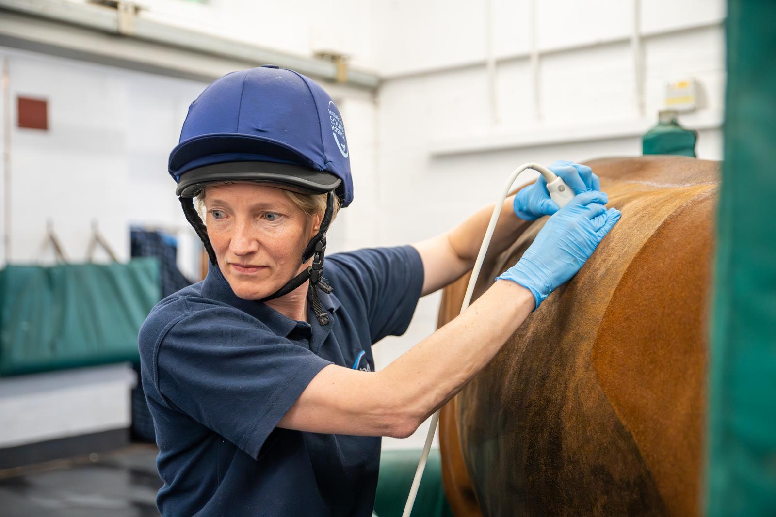
Ultrasound
We are equipped with an extensive variety of high-quality ultrasound scanners.
These are for use with our experienced ambulatory team to perform limb ultrasound, reproductive ultrasound and investigation of abdominal problems at the yard, and also in the hospital setting.
In the hospital we have larger cart-based powerful systems which can allow highly detailed tendon and ligament scanning, stifle and upper limb / pelvis imaging, thorough cardiac (heart) and lung imaging, and detailed investigation of abdominal problems such as colic, weight-loss and poor performance. We continually invest in the most up-to-date equipment to get the best for your horses, and our hospital specialists are available to discuss cases, receive second opinion referrals and examine emergencies 24 hours a day, 7 days a week, 365 days of the year.
Standing CT
In 2021, we became one of the first equine hospitals in the world to install a fully functioning standing cone beam CT unit for imaging of horses feet, pasterns and fetlock regions.
Cone-beam CT works in a slightly different way to CT performed under anaesthesia or that performed of the head, and is primarily considered beneficial in horses where a bony injury in the region of the foot to fetlock is considered most likely. This examination takes only 60 seconds to perform once the horse is positioned, and is done standing up, and is completely non-invasive. We occasionally complement the CT examination with use of CT contrast which can be injected into a joint (to look at articular cartilage).
Please note for imaging of the feet, the shoes will need to be removed, however this can be performed at the hospital if needed. Should a soft tissue injury be suspected, then MRI examination should be considered, as this is the best test for soft tissue imaging, we advise that individual cases are discussed with the imaging team at the hospital, who can advise on the most appropriate test for your individual horse – imaging@rainbowequinehospital.co.uk